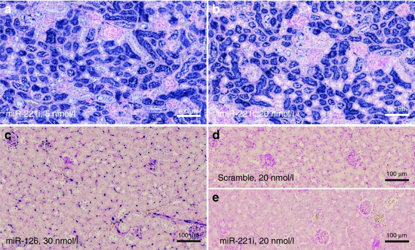
Figure 2.
In situ hybridization (ISH) analysis of LNA-i-miR-221. Serial sections from formalin-fixed paraffin-embedded (FFPE) tissue samples of NOD.SCID mouse kidney after a single i.p. (intraperitoneal) injection of 25 mg/kg LNA-i-miR-221 (a–d) or saline (e), processed by ISH with Locked Nucleic Acid (LNA) probes for LNA-i-miR-221 (a, b, e), miR-126 (c), and miR-221 scramble (d). Intense LNA-i-miR-221 ISH signal is detectable in tubules at both low (5 nmol/l) and high (20 nmol/l) probe concentrations (a, b), whereas the probe results in no ISH signal in saline-treated kidney. The positive control probe, miR-126, stains endothelial cells including capillaries in the glomeruli (c), and only background staining is seen by the use of the scramble negative control probe (d).

