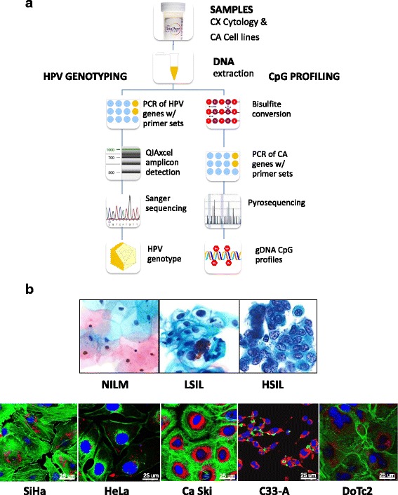
Fig. 1.

Protocol schema and representative images of cervical cytology and cervical carcinoma cell lines used in the study. a Sample collection, DNA extraction, HPV genotyping by Sanger sequencing, and CpG profiling of gene-specific promoters by pyrosequencing. b Three categories of liquid-based cervical cytology: negative for intraepithelial lesion or malignancy (NILM), low-grade squamous intraepithelial lesion (LSIL), and high-grade intraepithelial lesion (HSIL), reveal progressive nuclear enlargement, nuclear membrane irregularity, and chromatin coarseness associated with worsening grade. Five cervical carcinoma cell lines: SiHa, HeLa, Ca Ski, C33-A, and DoTc2, with distinct cytomorphologic features, e.g., cell size and shape, nucleus (blue), nuclear-to-cytoplasmic ratio, chromatin patterns, actin cytoskeleton (green), and mitochondria (red). Each cell line was immunofluorescence labeled and imaged by confocal microscopy (×63 objective). Abbreviations: CX cervical, CA cancer, PCR polymerase chain reaction, HSIL high-grade squamous intraepithelial lesion, LSIL low-grade squamous intraepithelial lesion, NILM negative for intraepithelial lesion or malignancy
