Fig. 1.

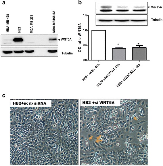
The loss of WNT5A induces “EMT-like” changes in human mammary epithelial HB2 cells. a Representative Western blot showing the presence of WNT5A protein in whole-cell lysates from MDA-MB468, HB2 and MDA-MB231 cells (n = 3). MDA-MB468 cells that were stably transfected with the WNT5A plasmid (MDA-MB468-5A) were used as a positive control for the experiment. Tubulin was used as a loading control. b Two different anti-WNT5A siRNA oligonucleotides were tested on HB2 cells. Total cell lysates were prepared 48 h after transfection, and Western blotting for the WNT5A protein was performed (as described in the Methods section). Error bars represent the standard error of the mean (n = 4). *p < 0.05. c Phase-contrast microscopy showing morphological changes in HB2 cells grown on glass coverslips and transfected with WNT5A siRNA for 48 h. The magnification used was 20X
