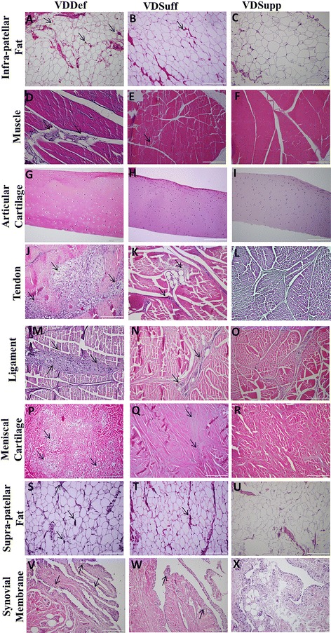
Fig. 1.

Hematoxylin and eosin (H&E) staining of infrapatellar fat, quadriceps muscle, articular cartilage, patellar tendon, collateral ligament, meniscal cartilage, suprapatellar fat, and synovial membrane. H&E staining shows the histology and inflammation in the vitamin D-deficient (VDDef), vitamin D-sufficient (VDSuff), and vitamin D-supplemented (VDSupp) group infrapatellar fat (a, b, c), muscle (d, e, f), articular cartilage (g, h, i), tendon (j, k, l), ligament (m, n, o), menisci (p, q, r), suprapatellar fat (s, t, u), and synovial membrane (v, w, x). Arrows indicate the presence of inflammatory cells in the tissue. These are the representative images of five swine each in the VDDef and VDSuff groups and three swine in the VDSupp group
