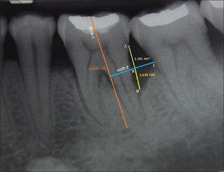
Figure 3.

Radiographic appearance at 9 months for site treated with PRF + DBM. AUX 1: Auxillary line 1 was drawn in the direction of tooth long axis, A: Cementoenamel junction (CEJ), C: Most coronal extension of the lateral wall of intrabony defect, AUX 2: Auxillary line 2 was drawn perpendicular to the tooth long axis and through point C, D: Base of the defect, B: Point where AUX 2 cross the contour of the root to point D
