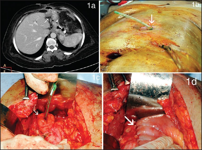
Figure 1.

(a) Cross section of contrast enhanced computed tomography scan scan showing a well-formed fistulous tract leading from the mid-sleeve to the midline laparotomy wound (b and c) Intraoperative photograph showing the gastro-cutaneous fistula opening at midline laparotomy scar with a nasogastric tube threaded across seen to arise from a large mid-body fistula at laparotomy (d) Intraoperative photograph showing the side-to-side fistula loop gastro-jujenostomy
