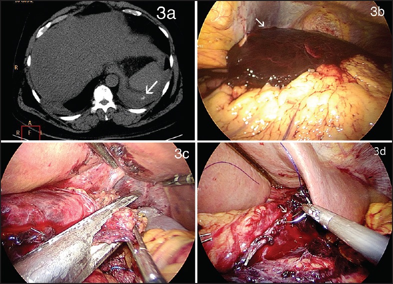
Figure 3.

(a) Cross section of contrast-enhanced CT scan revealed a sub-diaphragmatic collection suggestive of a haematoma with a small leak of contrast within the haematoma (b) Intraoperative view of staple line haematoma (c) Intraoperative photograph showing the stomach just beyond oesophago-gastric junction being re-sleeved with a linear stapler (d) Intraoperative photograph showing site of staple line bleeding managed by suture ligation
