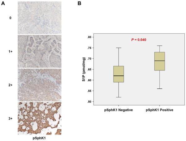
Fig. 3.
S1P levels are associated with phospho-sphingosine kinase 1 (pSphK1) expression. (A) Staining intensity is determined as shown; the staining intensity was scored as 0 (negative), 1 (weak), 2 (moderate), or 3 (strong). The staining score 0 and 1 was determined as pSphK1 negative and 2 and 3 determined as pSphK1 positive. (B) The levels of S1P were determined in the patients with pSphK1 negative or positive. Mean values are shown by horizontal lines. The statistical analysis was done by the Mann-Whitney U test.

