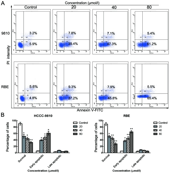
Figure 4.
Schisandrin B induces apoptosis in CCA cells. HCCC-9810 and RBE cells were treated with Sch B (0, 20, 40 and 80 µmol/l) for 48 h. (A) Flow cytometric analysis of apoptosis quantification by dual Annexin V-FITC and propidium iodide (PI) staining in untreated cells or Sch B-treated cells. (B) Percentages of surviving and early and late apoptotic cells are presented as the means ± SD (n=3). Significant differences from control experiments; *P<0.01, **P<0.001.

