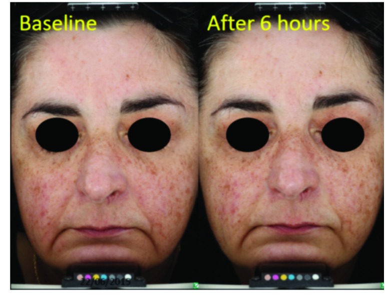
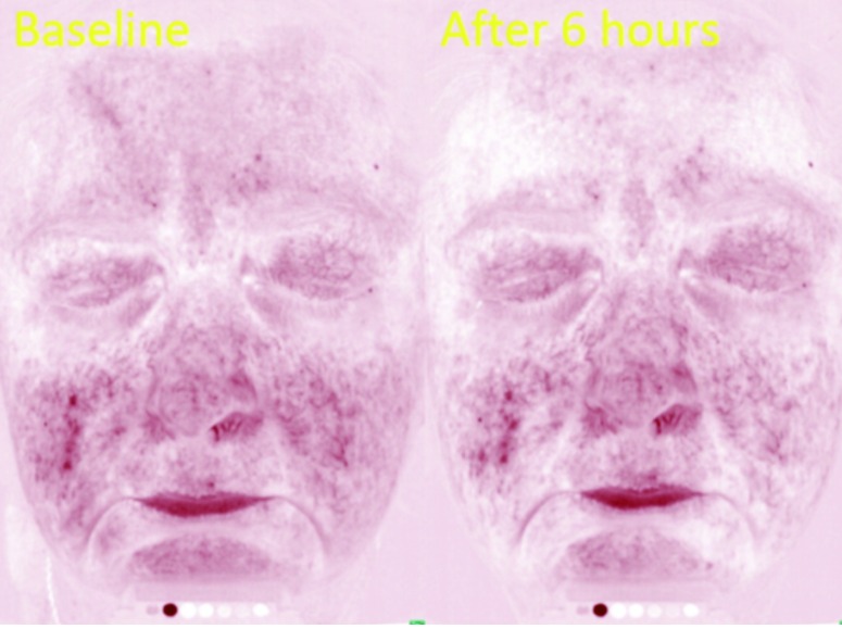
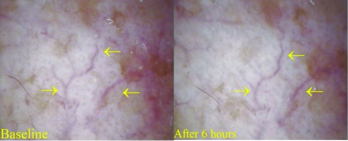
Figure 5.


Patient 1: Marked telangiectasia with minimal background erythema. (A) Polarized light photography and (B) erythema-directed photography using VISIA CR system (Canfield, US) of the patient at baseline and 6 hours after application of brimonidine 0.33% gel showing poor response to treatment; (C) ×30 videodermatoscopy at baseline and after 6 hours showing the persistence of telangiectasia (arrows). Videodermatoscopy is a tool enabling the magnification and measurement of vessels in patients with erythematotelangiectatic rosacea.

