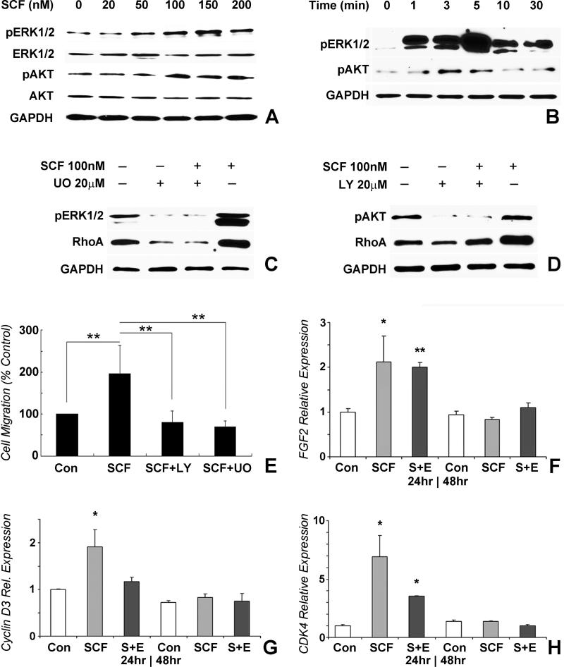
Figure 4. SCF modulated DP cell function through ERK and PI3K pathways.
DP cells were seeded into 6-well plates at 5×10⁵/well and cultured for pre-determined time intervals. SCF alone or SCF plus ERK/AKT inhibitor were then added to culture medium. (A) Western blot analysis of the dose-dependent effect of SCF on the phosphorylated state of ERK and AKT in DP cells. (B) Time course of the SCF effect on the phosphorylated state of ERK and AKT. (C) Inhibition of the SCF effect on ERK phosphorylation and RhoA protein expression by the ERK inhibitor. (D) Inhibition of the SCF effect on AKT phosphorylation and RhoA expression by the PI3K inhibitor. SCF function was selectively inhibited with the ERK inhibitor UO126, or the PI3K inhibitor LY 294002. (E) Blockage of the SCF effect on cell migration by both ERK and PI3K inhibitors. (F-H) Real-time RT-PCR analysis of cell proliferation related gene expression in dental pulp progenitors treated with SCF only or SCF (SCF) plus ERK inhibitor (S+E) compared to controls (Con) and after 24 or 48 hours cell culture. Levels of gene expression of FGF2 (F), Cyclin D3 (G), and CDK4 (H) were calculated relative to control group gene expression levels after 24 and 48 hours of culture. * p<0.05, ** p<0.01.

