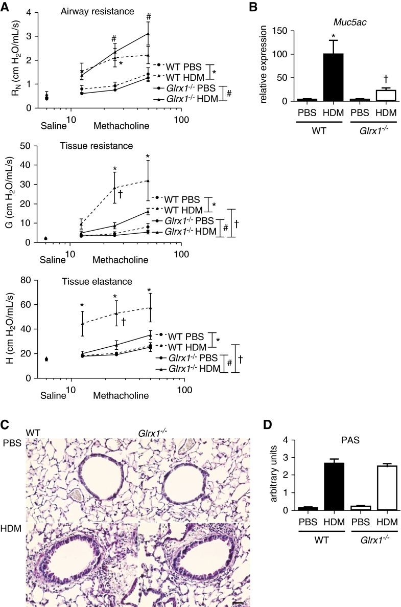
Figure 6.
Evaluation of alterations in respiratory mechanics and mucus metaplasia in WT and Glrx1−/− mice in response to 15 challenges of 50 μg of HDM. (A) Assessment of airway hyperresponsiveness (AHR) via a forced oscillation technique in WT and Glrx1−/− mice. Changes in respiratory mechanics were analyzed 72 hours after the final instillation of either PBS or HDM. Ascending doses of methacholine were administered to determine Newtonian resistance (RN), tissue resistance (G), and tissue elastance (H). Data are expressed as means (± SEM; six to roughly eight mice per group). For AHR data, a three-way ANOVA was used with an additional term for methacholine dose. */#P < 0.05 compared with PBS controls; †P < 0.05 compared with WT mice challenged with HDM. (B) Evaluation of mucin-5AC (Muc5ac) gene expression in homogenized lung tissue. mRNA was analyzed by real-time RT-PCR, and results were normalized to the housekeeping gene cyclophilin and are expressed as fold increases in expression compared with WT PBS controls. Data are expressed as means (± SEM; six mice per group). *P < 0.05 (ANOVA) compared with PBS controls; †P < 0.05 (ANOVA) compared with WT mice challenged with HDM. (C) Periodic acid–Schiff (PAS) staining of airway mucus in WT or Glrx1−/− mice exposed to HDM or PBS (scale bar, 50 μm). (D) Quantification of airway mucus staining (PAS) intensity was determined by scoring of slides by two blinded investigators. Data are expressed as means (± SEM) from six to eight mice per group. *P < 0.05 (Kruskal–Wallis) compared with respective PBS control groups. †P < 0.05 (Kruskal–Wallis) compared with WT HDM groups.

