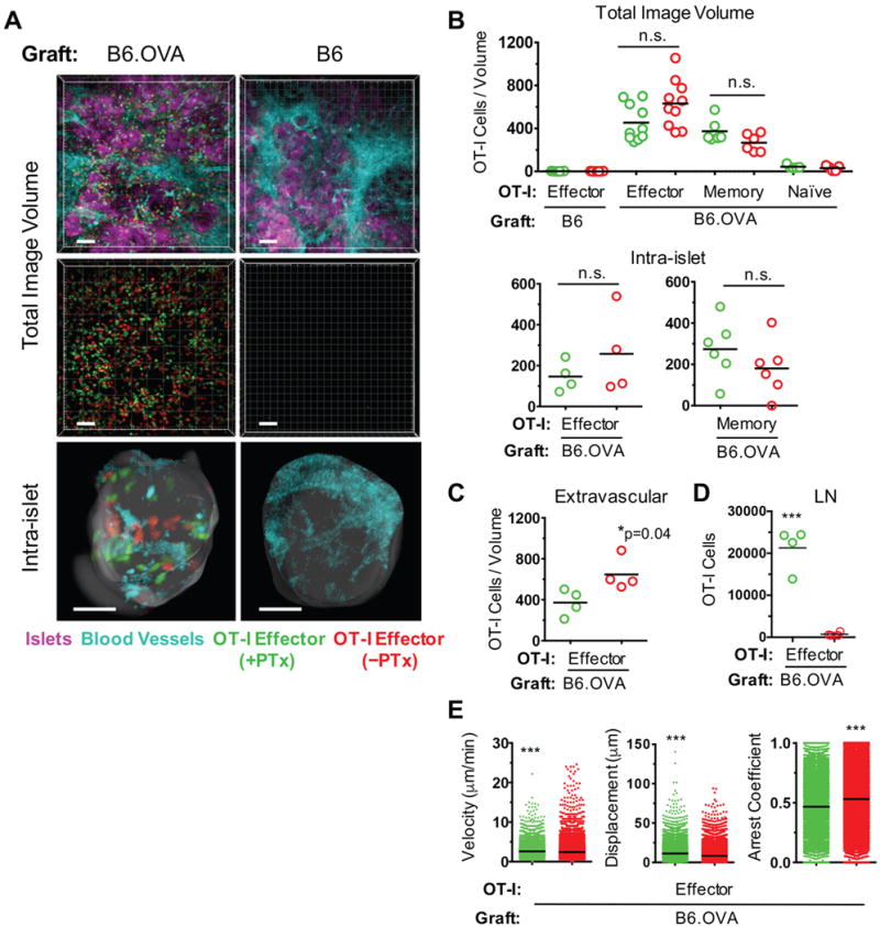
FIGURE 1. Role of cognate Ag and Gαi-coupled chemokine receptor signaling in CD8+ effector T cell migration.

B6 Act-OVA transgenic (B6.OVA) or non-transgenic B6 islets were transplanted under the kidney capsule of diabetic B6 recipients. PTx-treated (green) and untreated (red) OT-I effector, naïve, or memory T cells were co-transferred 9 – 10 days after transplantation and imaged by two-photon intravital microscopy 16 – 24 hr later. All OT-I cells were from B6 OT1.RAG−/− donors. (A) Representative, volume-rendered images depicting PTx-treated and untreated OT-I effectors present in either the total or intra-islet image volumes. Blood vessel and islet channels were subtracted from middle panels to better visualize infiltrating T cells. Scale bar = 50 μm. (B) Enumeration of OT-I cells detected in total and intra-islet image volumes. Each data point represents number of cells in one movie (image volume). For OT-I effector migration, 5 mice in the B6.OVA group and 3 mice in the B6 group were imaged. For analysis of naïve and memory OT-I migration, 2 mice were imaged in each group. (C) Enumeration of extravascular OT-I effectors in the total image volume. (D) Enumeration of OT-I effectors in lymph nodes of recipient mice. (E) Motility parameters of OT-I effectors in total image volume. *p<0.05; ***p<0.0001; n.s. = not significant.
