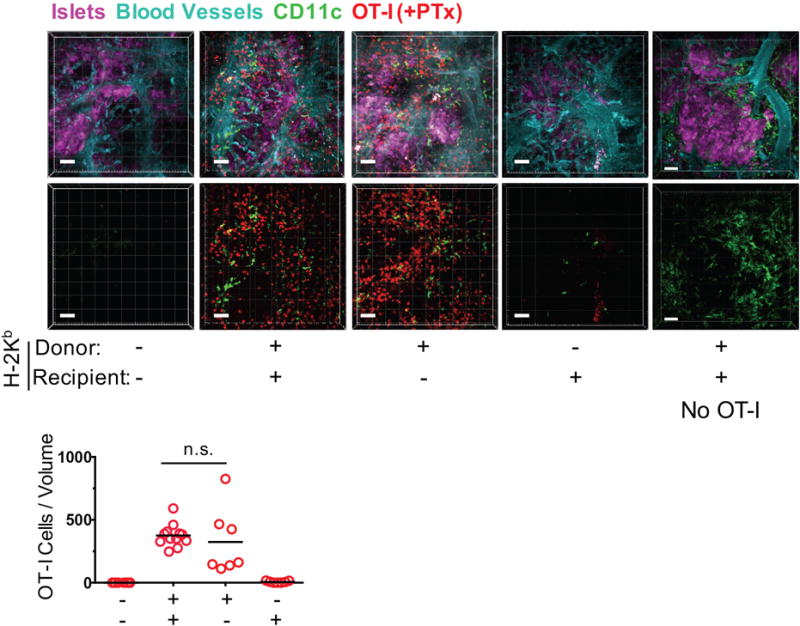
FIGURE 2. Cognate Ag presentation by donor graft cells is necessary for CD8+ effector T cell migration.

Migration of PTx-treated OT-I effector T cells to B6.OVA islet grafts (in which recipient cells (N = 5 mice, 7 movies), donor cells (N = 3 mice, 7 movies), or both (N = 2 mice, 8 movies) lacked the H-2Kb MHC class I molecule required for presenting OVA peptide to OT-I cells) was imaged and enumerated by two-photon intravital microscopy as described in Fig. 1, except that recipients also carried the YFP transgene under control of the CD11c promoter to visualize recipient-derived DC in the graft. Grafts in which both donor and recipient cells expressed H-2Kb served as positive controls (N = 4 mice, 12 movies). Additional grafts in which both donor and recipient cells expressed H-2Kb but no OT-I cells were transferred were also imaged (far right panels; N = 2 mice, 10 movies). Representative volume-rendered images are shown. Blood vessel and islet channels were subtracted from bottom panels to better visualize infiltrating T cells. n.s. = not significant. Scale bar = 50 μm.
