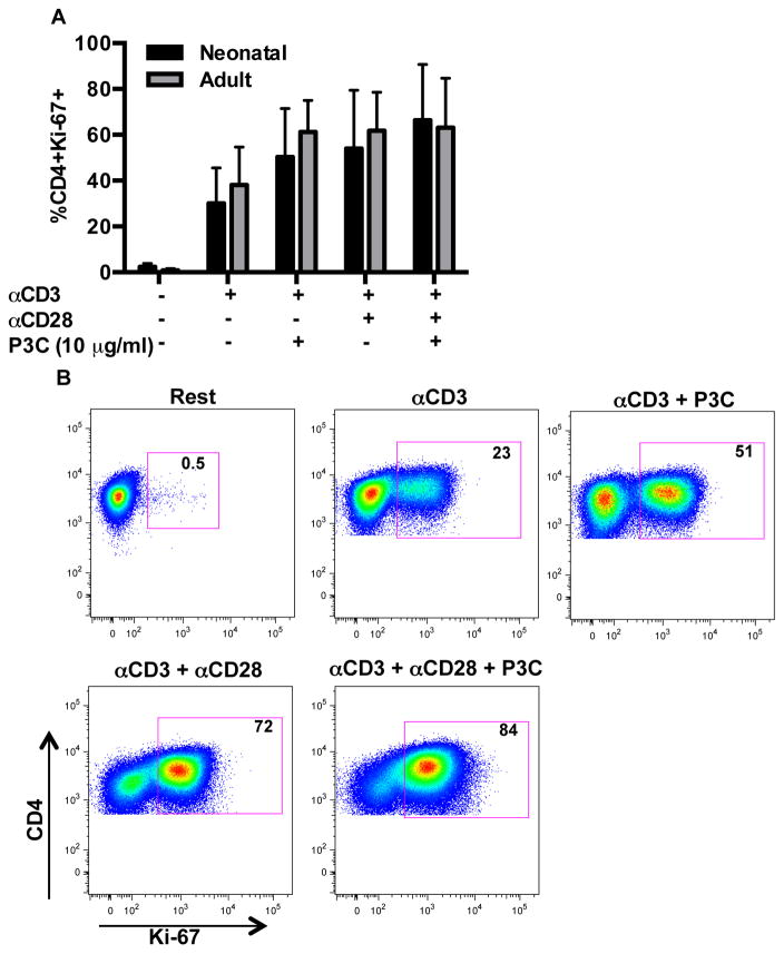
Figure 8. TLR-2 induced CD4+ T cell proliferation in neonatal and adult donors.
To assess for T cell proliferation, purified CD4+CD45RA+ T cells were stimulated as indicated in 24 well tissue culture plates for 96 h and subsequently stained with LIVE/DEAD Aqua and CD4-PECy7, followed by fixation and permeabilization in FoxP3 staining buffer and staining with anti-Ki-67-BV421. (A) Shown are mean percentages of viable CD4+ T cells with Ki-67 expression ± SD (n=7 cord; n=7 adult). Data were analyzed using two-way ANOVA followed by Tukey-Kramer adjustment for multiple comparisons. Adjusted p values < 0.05 were considered significant. Minimal increases in Ki-67 expression were observed with addition of P3C to αCD3 ± αCD28 in both age groups (p > 0.05 all comparisons). The proliferative response of neonatal and adult naïve CD4+ T cells did not differ significantly under any of the tested conditions.
(B) A representative flow plot from a neonatal donor is shown.

