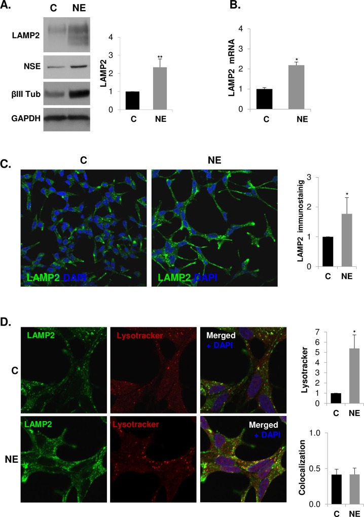
Fig 3. Lysosomal-associated membrane protein 2 (LAMP2) is over-expressed in neuroendocrine differentiated LNCaP cells.
LNCaP cells were grown in serum-containing medium (C, control cells) or serum-free medium (NE, neuroendocrine cells) during 6 days. (A) LAMP2 was measured in whole lysates of C and NE cells by western blot along with neuroendocrine markers neuron-specific enolase (NSE) and βIII tubulin (βIII Tub). GAPDH was used as a loading control. Densitometric analysis of the Western blot bands are shown on the right. (B) Quantification of LAMP2 mRNA in control and NE cells by real time qRT-PCR. Results are the mean ± S.D. of at least three independent experiments (* p<0.05 versus control cells compared by the Student’s t test). (C) Detection of LAMP2 by immunofluorescence (green). Nuclei are stained with DAPI (blue). Immunofluorescence was analyzed by confocal microscopy. Quantitative analysis of lysotracker and LAMP2 was performed using ImageJ software (NIH).

