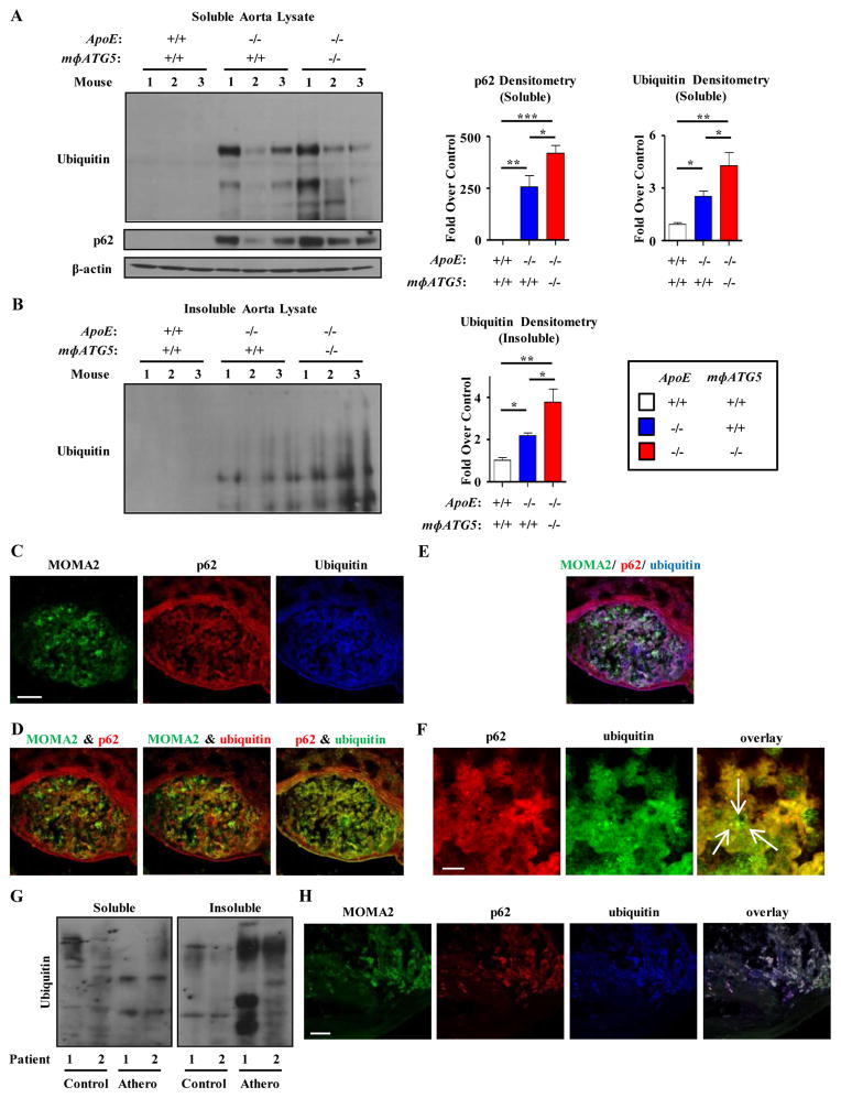
Figure 2. The accumulation of insoluble p62-enriched polyubiquitinated proteins in macrophages is characteristic of both mouse and human atherosclerosis.
(a–b) Western blot analysis of polyubiquitinated proteins and p62 in soluble (a) and insoluble (b) lysates of whole aortas from control (WT), ApoE−/−, and ApoE−/−/mΦATG5−/− mice (n=3 mice for each genotype) fed a Western Diet for 2 months. Densitometric quantification and a representative western blot are shown. (c–f) Representative immunofluorescence images of aortic roots from ApoE−/− mice co-stained with antibodies against the macrophage marker (MOMA2), ubiquitinated proteins (FK-1 antibody), and p62. Individual stains (c), overlays of 2 stains (d), overlays of all 3 stains (e), and magnified views of areas of punctate co-localization between p62 and Ubiquitin (f) are shown. Arrows indicate dots co-stained for both ubiquitin and p62. Images are representative of eight independent experiments. (g) Western blot analysis of polyubiquitinated proteins in soluble and insoluble lysates of atherosclerotic plaques and adjacent non-atherosclerotic regions extracted from post-operative human carotid endarterectomy specimens. (h) Immunofluorescence images of an atherosclerotic region from a carotid endarterectomy specimen co-stained with MOMA2, ubiquitinated proteins (FK-1 antibody), and p62. Images are representative of two carotid endarterectomy specimens. For all graphs, data are presented as mean ± SEM. *P<0.05, **P<0.01, ***P<0.001. Scale bar, 100 μm (c,h); 10 μm (f).

