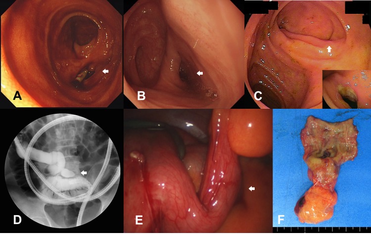
Fig 1.
Meckel’s diverticulum (A-C) Balloon-assisted enteroscopy showed double-lumen signs and/or concurrent ulcerative lesions in the lumens (D) During balloon-assisted enteroscopy, about 4 cm sized luminal out-pouching diverticulum was identified in the distal ileum under fluoroscopy after radio-contrast dye injection, (E) During laparoscopy, about 4 cm sized diverticulum was identified, and (F) Surgical specimen of small bowel resection and anastomosis for Meckel’s diverticulum.

