
Fig 6. Effects of forced TFF3 expression on RB cell migration.
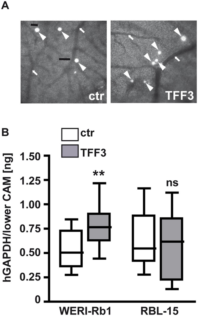
(A) Grayscale pictures (100x) depicting GFP-labeled WERI-Rb1 cells (arrowheads), which exited the vasculature (arrows) after injection into a CAM vein. (B) Quantification of hGAPDH content (normalized against 18S RNA) in lower CAM punches 7 days after intravenous injection of TFF3 overexpressing (TFF3) and control (ctr) WERI-Rb1 and RBL-15 cells. Values are means from at least 3 independent experiments ± SEM.; **P < 0.01; ns = no statistical differences compared to the control group calculated by Student`s t-test.