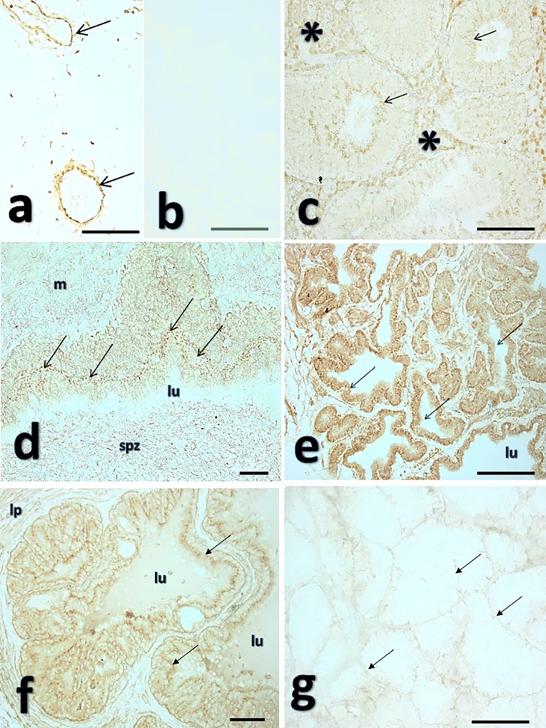
Fig 4. Immunolocalization of glutathione peroxidase-5 (GPX5) in boar genital organs.
Fig 4a depicts a representative positive control section depicting immunostained blood vessels with a major restricted endothelial GPX-5 localization (arrows). A representative negative control prostate section (primary Ab omitted) is depicted in Fig 4b. Fig 4c shows a representative section of boar testis, showing immunostaining in the interstitium (blood vessels and Leydig cells, asterisk) and the seminiferous tubules, with a marked immunostaining in the elongated spermatids (arrows). The immunostaining was more marked in the lining epithelium of the ductus epididymides (Fig 4d, cauda segment) where both the principal epithelial cells (arrows point to the apical cell region, base of the stereocilia) and the luminal spermatozoa (spz) appeared stained, while the surrounding smooth muscle appears only faibly stained (m), lu: lumen. Fig 4e-g depict accessory sexual glands secretory epithelia of the prostate (4e), the seminal vesicles (4f) and the bulbourethral gland (4g). While prostate depicted both nuclear and cytoplasmic staining (4e), the seminal vesicles depicted mainly cytoplasmic staining (arrows) and a clear staining in the luminal secretion (lu), lp: lamina propria. The bulbourethral gland (4g) was immunostained in the blood vessel dominated interstitial with some staining in the latero-basal epithelial membrane (arrows). Not-counterstained sections, viewed with phase-contrast optics. Bars: 50μm.

