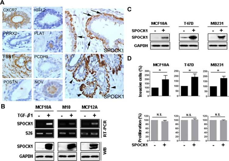
Fig 1. SPOCK1 was a TGF-β-induced myoepithelial marker and SPOCK1 overexpression enhanced invasiveness.
(A) Immunohistochemistry demonstrated that SPOCK1 was the only myoepithelial marker among the evaluated TGF-β–upregulated gene products in MCF10A cells. CXCR7 staining was observed in luminal epithelia but not myoepithelia whereas FBN1 staining was observed diffusely in the stroma but not in the epithelia. IGFL2, PRRX2, PLAT, PCDH9, POSTN and NOV staining was not found in the ductolobular units (left and middle panels). By contrast, SPOCK1 was immunolocalized within (arrow) or beneath (arrowhead) the myoepithelia (right panel). (Magnification × 400). (B) Upregulation of SPOCK1 at mRNA levels (upper panels) and protein levels (lower panels) were observed in MCF10A, M10, and MCF12A cells, 3 days after treatment with TGF-β. S26 was used as an mRNA loading control and GAPDH was used as a protein loading control. (C) Western blotting was used to detect the expression of SPOCK1 in MCF10A, T47D, and MB231 cells 24 h after transient transfection with pCDH-SPOCK1 or control vector pCDH. (D) The invasive capability and proliferation were measured in the cells shown in (C). Data from invasion assay are shown as the mean ± SD of 3 fields. Data from MTT assay are shown as mean ± SD of 3 independent experiments. These results are presented as the percentage relative to their control cells (*, P < 0.05; N.S., nonsignificant).

