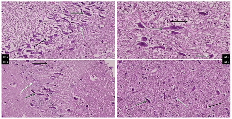
Fig 6. Brain lesion evaluations.
Brain lesions were assessed in short bowel rats administered diclofenac (D, 12 mg/kg), saline 5 mL/kg, or BPC 157 (10 μg/kg) intraperitoneally. Characteristic presentation of hippocampal (HC and HB) and cerebellar nuclear (CC and CB) lesions are shown. Left upper panel (control, HC) shows severe edema and neuronal damage (gray arrow indicates ballooned; black, edematous; and white, normal neurons); right upper panel (BPC 157, HB) shows less edema and neuronal damage (gray arrow indicates ballooned; black, edematous; and white normal neurons). Cerebellar nuclei, lower panel (control (HC) shows severe edema and neuronal damage (gray arrow indicates ballooned; black, edematous; and white normal neurons); right lower (BPC 157, HB) shows less edema and neuronal damage (gray arrow indicates ballooned; black, edema; and white, normal neurons), H&E, 40×.

