
Fig. 3.
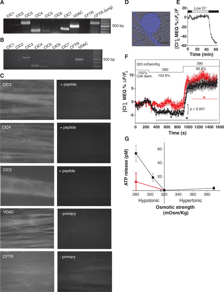
VAACs in DRG neurons. (A) RT-PCR analysis detected mRNA for several chloride channels but not CFTR. mRNA from lung is a positive control. (B) RT-PCR from neurons collected individually from cell cultures detected only ClC3, ClC4, ClC5, and VDAC. (C) Positive immunocytochemical staining with antibodies against ClC3, ClC4, ClC5, and VDAC was found in DRG axons; CFTR immunostaining was negative. No staining was seen when antibody was preabsorbed with peptide (+ peptide) or in the absence of primary antibody (− primary). (D) Functional VAACs in DRG neurons are shown by monitoring changes in cytoplasmic Cl− by two-photon microscopy with the fluorescence indicator MEQ (blue fluorescence superimposed on DIC image of DRG cell body). (E) Influx of Cl− quenches MEQ fluorescence as shown by perfusing cells in normal saline after incubation in low Cl− saline, in which 145 mM sodium gluconate was substituted for NaCl. (F) VAAC function is shown by the influx of Cl− during cell swelling (decreased MEQ fluorescence, black trace) produced by switching from isotonic (320 mOsm/kg) to hypotonic conditions (280 mOsm/kg), and efflux of Cl− is seen during cell volume reduction in 390 mOsm/kg solution. Cl− influx during cell swelling was significantly inhibited by 100 μM NPPB (red) (−3.56 ± 0.12 versus −1.09 ± 0.074; P < 0.001). Asterisks indicate when responses are significantly different from baseline in the presence (red) or absence (black) of NPPB. ANOVA and Dunnett’s post hoc analysis of responses compared at sequential intervals of 200 s, relative to isotonic solution (320 mOsm/kg). Mean and SEM of responses in 11 neurons are plotted. Changes in cell diameter are plotted as percentage relative to starting conditions in isotonic saline (defined as 100%). (G) Hypotonic saline below the normal osmotic strength of 320 mOsm/kg caused release of ATP from DRG neurons in the absence of electrical stimulation. ATP release was significantly inhibited by the chloride channel blocker, 100 μM NPPB (red triangle) (n = 12), indicating release through VAACs (P < 0.001, ANOVA, n = 56). Scale bar, 20 μm; 100-bp cDNA ladder in (A) and (B).