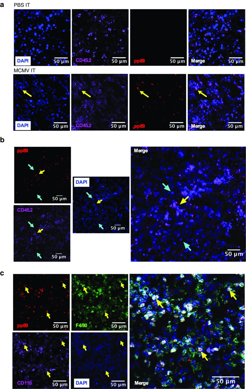
Figure 4.
MCMV infects TAMs after IT therapy. Mice received IT injections with WT-MCMV or MCMV-gp100 as in Figure 3. Tumors were harvested 1 day after the last IT injection and processed for histology. Yellow arrows indicate pp89 positive cells. Cyan arrows indicate pp89 negative cells. (a,b) Immunofluorescence staining of pp89 (red) in tumors IT injected with PBS or MCMV. Tumors were also stained for hematopoietic cells (CD45.2, purple) and costained with DAPI (blue). (c) pp89 (red) expression colocalizes with macrophages expressing CD11b (purple) and F4/80 (green) cells, after MCMV IT infection. CMV, cytomegalovirus; MCMV, vaccination with murine-CMV; PBS, phosphate-buffered saline; DAPI, ; TAMs, tumor associated macrophages; IT, intratumoral.

