Figure 6.
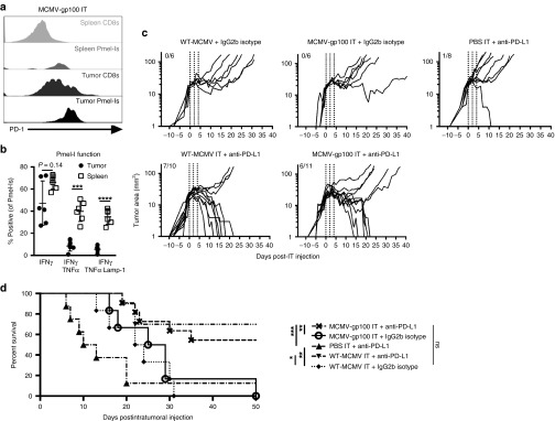
IT MCMV treatment combined with anti-PD-L1 therapy profoundly improves B16F0 tumor growth delay and survival. For a and b, Mice received 1 × 10⁴ Pmel-Is one day prior to tumor implantation and were IT infected with MCMV as in Figure 3. (a) Representative histograms of the PD-1 expression of CD8+ T-cells or Pmel-Is 7 days post infection. (b) Ex vivo cytokine production and degranulation in response to native gp100 stimulation of Pmel-Is 7 days post infection and represented as the mean ± SD. Significance was assessed by a paired t-test, *P < 0.05; **P < 0.01; ***P < 0.001; ****P < 0.0001. (c) Mice bearing B16F0 tumors were treated with anti-PD-L1 or an isotype control antibody beginning on the day of MCMV IT infection. Shown is the tumor growth as in Figure 3 for the indicated groups of mice. Vertical dotted lines represent days of MCMV IT infection. Fractions in each graph represent the number of animals that cleared the tumor out of the number of animals tested. (d) Kaplan–Meier survival curve of the mice in each treatment group. Significance was assessed by a logrank test, P > 0.05 is nonsignificant; *P < 0.05; **P < 0.01; ***P < 0.001; ****P < 0.0001. CMV, cytomegalovirus; MCMV, vaccination with murine-CMV; PBS, phosphate-buffered saline; IT, intratumoral.

