Figure 5.
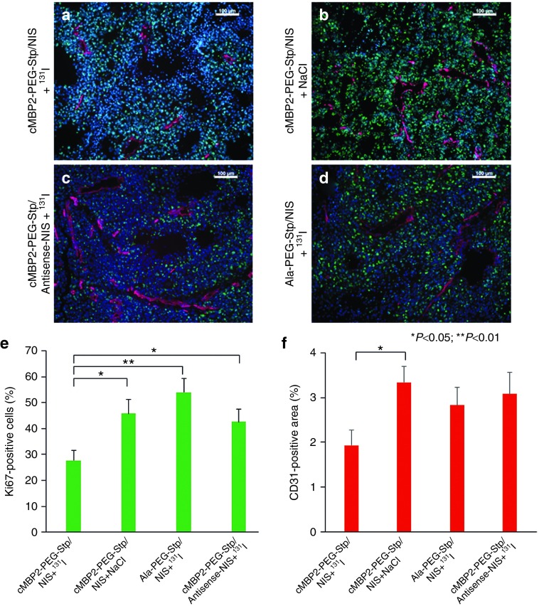
Immunofluorescence analysis. After treatment, when tumors reached a size of 1,500 mm3, mice were sacrificed and tumors dissected. Frozen sections of tumor tissue were stained with a Ki67-specific antibody (green) to determine cell proliferation and an antibody against CD31 (red) to label blood vessels (a–d). Tumor cell proliferation (e) and blood vessel density (f) in tumors from animals treated with cMBP2-PEG-Stp/NIS that received 131I (n = 10) were compared to control groups treated with cMBP2-PEG-Stp/NIS+NaCl (n = 8), Ala-PEG-Stp/NIS+131I (n = 6) or cMBP2-PEG-Stp/Antisense-NIS+131I (n = 6). Nuclei were counterstained with Hoechst. Results are reported as mean ± standard error of the mean. Scale bar = 100 µm.

