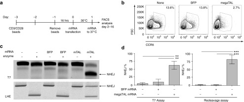
Figure 3.
megaTAL efficiently disrupts CCR5 in primary CD4+ cells. (a) Timeline representing workflow in primary CD4+ cells relative to time of transfection (t = 0), beginning with the addition of anti-CD3/CD28 beads on cryo-preserved CD4+ T cells. (b) Surface staining of CCR5 in primary CD4+ cells comparing expression in untreated cells or cells transfected with BFP or CCR5 megaTAL mRNA. (c) Representative agarose gels quantifying CCR5 modification by T7 endonuclease assay (surveyor assay; top panel) or by re-cleavage assay (RCA) using a fluorescently labeled forward primer (lower panel). (d) Molecular quantification (n = 3) by T7 (left) or RCA (right) of CCR5 disruption in primary CD4+ cells. Values are calculated from fluorescent densitometry (%NHEJ = NHEJ band/sum of NHEJ + undisrupted bands).

