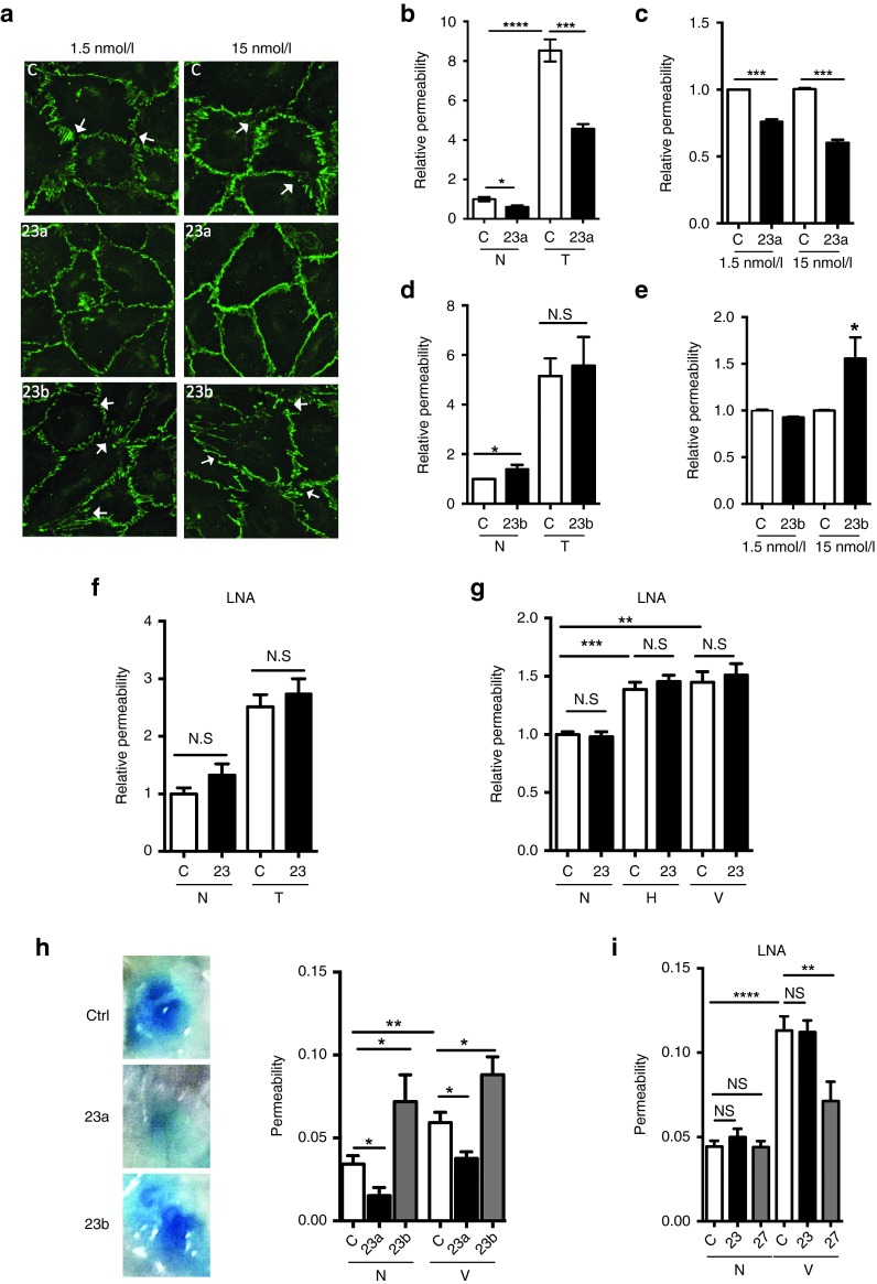
Figure 4.
MiR-23a inhibits and miR-23b enhances EC permeability. Cells were stimulated with 0.1–0.2 U/ml of thrombin, 10 µmol/l of histamine or 50 ng/ml of human VEGF165 for 30 minutes as indicated for in vitro experiments. (a) 48 hours after 1.5 or 15 nmol/l of miR-23a or miR-23b mimic treatment, HUVEC were stained for VE-cadherin (green). White arrows = examples of changed adherens junctions (VE-cadherin-staining). (b,d) Permeability measured in control or miR-23a (b) or miR-23b (d)-mimic–transfected cells without (N) or with thrombin stimulation (T). n = 4 experiments. (c,e) Permeability measured at basal level in control or cells transfected with 1.5 or 15 nmol/l miR-23a-mimic (c) or miR-23b (e). n = 3 experiments, where values are normalized to control (C). (f,g) Permeability was measured without (N) or with thrombin (T, n = 6 experiments), histamine (H, n = 4 experiments) or VEGF (V, n = 5 experiments) stimulation in control or miR-23 LNA transfected EC. (h) The Miles assay was performed with 4 µg of control mimic or miR-23a or miR-23b mimic injected intradermally into the back of the mice. 24 hours later, 200 μl 0.5% Evan's Blue was injected intravenously. After 30 minutes, 10 ng VEGF (V) or phosphate-buffered saline (N) was given into the same site as the mimic. Mice were sacrificed 30 minutes later and the dye was extracted from skin samples and quantified; # mice are: N+C, n = 10; N+23a, n = 6; N+23b, n = 5; V+C, n = 12, V+23a, n = 11; V+23b, n = 6. Representative photos of lesions are given. (i) The Miles assay was performed with 4 µg of control LNA, LNA-23 or LNA-27 without (N) or with VEGF stimulation (V) with set up given as in f above. # mice are: N+C, n = 16; N+23, n = 16; N+27, n = 8; V+C, n = 16, V+23, n = 16; V+27, n = 8. All graph represents mean ± SEM; *P < 0.05; **P < 0.01; ***P < 0.001, ****P < 0.0001 by unpaired two-tailed t-test. LNA, locked nucleic acid.

