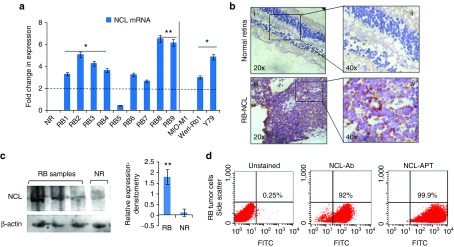
Figure 1.
Expression of nucleolin in RB tumor samples and cell lines. (a) Fold changes in gene expression of NCL in various primary cells and cell lines. (b) Immunohistochemistry of the normal retina section (i, ii), RB tissue sections (iii, iv). (c) Expression of NCL in the cytoplasmic fraction of RB tumor tissues by immunoblotting for NCL and β-actin, graph on the right shows the densitometry analysis of tumor samples normalized to normal retina (NR). (d) Scatter plots show the expression of NCL and the NCL-APT binding to RB tumor (RB cells from the enucleated eyes). The error bar represents the SD and the * indicates significance of P < 0.05 and ** indicates significance of P < 0.001 when compared to the normal retina.

