
Figure 1.
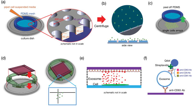
Single-cell assay used for analyzing exosome secretion. (a) Loading single cells onto a culture plate utilizing a PDMS mesh which can be removed afterward. (b) The mesh was removed after cell attachment. (c) A surface functionalized glass slide was placed on the support frame 100 μm above the cells. (d) and (e) The glass slide collected exosomes secreted by the corresponding cells. The fiducials on the cover glass served as registration marks. (f) The captured exosomes were labeled with another biotinylated antibody and streptavidin-conjugated Quantum dots to become visible under fluorescent microscope.