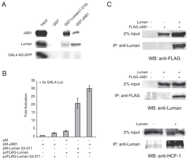
Fig. 1.
Luman interacts with JAB1 both in vitro and in vivo. (A) Direct binding of Luman to JAB1 in GST pull-down assays. GST, GST-Luman(1–215) and GST-JAB1 proteins were coupled to glutathione-sepharose beads and incubated with [35S]-labeled GAL4 AD-GFP, FLAG-Luman and HA-JAB1. After extensive washing, proteins were eluted from the beads, separated on a 10% SDS-PAGE gel and visualized by autoradiography. The input lanes have 10% of the radio-labeled protein used in each pull-down assay. (B) Luman interacts with JAB1 in the cell as demonstrated by the mammalian two-hybrid assay. HEK-293 cells were transiently transfected with a combination of pM-JAB1 and a Luman construct, along with the reporter plasmids p5×GAL4 luciferase and pRL-SV40. Dual luciferase activities were measured 24 h post-transfection. Values are normalized to Renilla luciferase before being referenced to the pM-JAB1 background control. Data are based on 6 independent assays and shown with standard errors. (C) Luman interacts with JAB1 in mammalian cells as demonstrated by co-immunoprecipitation. HEK-293 cells were transiently transfected with pcDNA-Luman, pFLAG-JAB1 or both. Cells were treated with proteasome inhibitor MG132 for the last 6 h, prior to harvest of cell lysates at 24 h post-transfection. Cleared lysates were incubated with either a Luman- or FLAG-specific antibody, followed by precipitation with Protein G beads. Precipitated samples were subjected to SDS-PAGE and probed in Western blotting with antibodies against FLAG (top panel), Luman (middle panel) or HCF-1 (bottom panel). HCF-1 was included as a positive control for Luman since it is a known interacting protein. The input lanes have 2% of cleared cell lysates used in the immunoprecipitation assays. Abbreviations: IP, immunoprecipitation antibody; WB, Western blot antibody.

