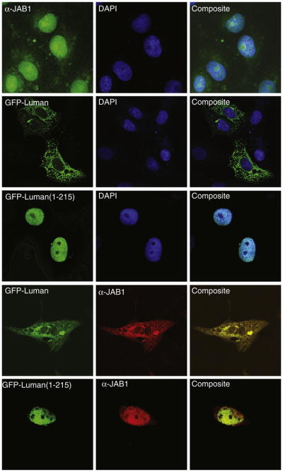
Fig. 2.
Co-localization of JAB1 with Luman demonstrated by confocal fluorescence microscopy. Vero cells were transiently transfected with plasmids expressing JAB1 or GFP-Luman protein. JAB1 was visualized indirectly with a JAB1-specific antibody and a fluorophore Alexa488- or Alexa594-conjugated secondary antibody. DAPI was used to counter-stain the nuclei of the cells.

