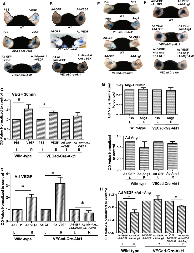
Fig. 3.
Akt1 augments VEGF-induced vascular permeability and Ang-1-induced vascular-barrier protection in vivo. a Representative images of PBS and 30 µl of 20 ng/ml recombinant VEGF administered WT mice (top), tamoxifen-treated VECad-Cre-Akt1 mice administered with PBS and VEGF (middle) and tamoxifen-treated VECad-Cre-Akt1 mice administered with Ad-GFP + VEGF and Ad-MyrAkt1 + VEGF ears for 30 min (short term) showing leakage of Evans blue dye (Miles assay). b Representative images of Ad-GFP and Ad-VEGF administered WT mice (top), tamoxifen-treated VECad-Cre-Akt1 mice administered with Ad-GFP and Ad-VEGF (middle) and tamoxifen-treated VECad-Cre-Akt1 mice ears expressing either Ad-GFP or Ad-myrAkt1 in the absence or presence of Ad-VEGF expression (long-term VEGF treatment) (bottom), showing leakage of Evans blue dye (Miles assay). c Histogram showing calorimetric quantification of the extravasated dye in the ears from PBS and 30 µl of 20 ng/ml recombinant VEGF administered WT mice (top), tamoxifen-treated VECad-Cre-Akt1 mice administered with PBS and VEGF (middle) and tamoxifen-treated VECad-Cre-Akt1 mice administered with Ad-GFP + VEGF and Ad-MyrAkt1 + VEGF after 30 min (n = 6). d Histogram showing calorimetric quantification of the extravasated dye in the ears from Ad-GFP and Ad-VEGF administered WT mice (top), tamoxifen-treated VECad-Cre-Akt1 mice administered with Ad-GFP and Ad-VEGF (middle) and tamoxifen-treated VECad-Cre-Akt1 mice ears expressing either Ad-GFP or Ad-myrAkt1 in the absence or presence of Ad-VEGF expression (long-term VEGF treatment) (n = 6). e Representative images of PBS and 30 µl of 50 ng/ml recombinant Ang1 administered WT mice and tamoxifen-treated VECad-Cre-Akt1 mice ears administered with PBS and 30 µl of 50 ng/ml recombinant Ang1 for 30 min (short term) (top two panels), and representative images of mice ears expressing Ad-GFP or Ad-Ang-1 (long term) in WT tamoxifen-treated VECad-Cre-Akt1 mice (bottom two panels) showing leakage of Evans blue dye (Miles assay). f Representative images of WT (top) and tamoxifen-treated VECad-Cre-Akt1 (middle) mice ears administered with combinations of either Ad-GFP/Ad-VEGF or Ad-VEGF/Ad-Ang-1, as well as VECad-Cre-Akt1 mice ears administered with a combination of either Ad-GFP/Ad-VEGF/Ad-Ang-1 or Ad-myrAkt1/Ad-VEGF/Ad-Ang-1 (bottom), showing leakage of Evans blue dye (Miles assay). g Histogram showing calorimetric quantification of the extravasated dye in the ears from WT and tamoxifen-treated VECad-Cre-Akt1 mice ears, with PBS or 30 µl PBS containing 50 ng/ml recombinant Ang-1 for 30 min, as well as WT and VECad-Cre-Akt1 mice ears, expressing either Ad-GFP or Ad-Ang-1 (n = 6). h Histogram showing calorimetric quantification of the extravasated dye in the ears from WT and tamoxifen-treated VECad-Cre-Akt1 mice administered with combinations of either Ad-GFP/Ad-VEGF or Ad-VEGF/Ad-Ang-1, as well as tamoxifen-treated VECad-Cre-Akt1 mice ears administered with a combination of either Ad-GFP/Ad-VEGF/Ad-Ang-1 or Ad-myrAkt1/Ad-VEGF/Ad-Ang-1 (n = 6). # P < 0.05, *P < 0.01

