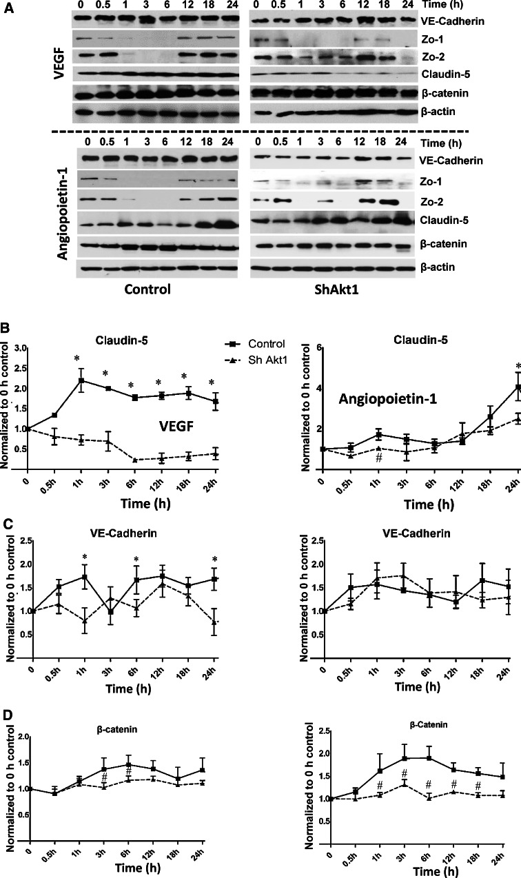
Fig. 4.
Akt1 deficiency affects real-time changes in the expression of proteins in endothelial-barrier AJs and TJs in response to VEGF and Ang-1 treatments. a Representative western blot images of control (left) and ShAkt1 (right) HMEC lysates treated with 20 ng/ml VEGF and real-time changes in the expression levels of AJ proteins VE-Cadherin and β-catenin as well as TJ proteins Zo-1, Zo-2 and claudin-5 were determined, and compared to 0 h time point (above; n = 3). Representative western blot images of control (left) and ShAkt1 (right) HMEC lysates treated with 50 ng/ml Ang-1 and real-time changes in the expression levels of AJ proteins VE-cadherin and β-catenin as well as TJ proteins Zo-1, Zo-2 and claudin-5 were determined, and compared to 0 h time point (below; n = 3). b Densitometry analysis of Western blot images of control and ShAkt1 HMEC lysates treated with 20 ng/ml VEGF (left) and 50 ng/ml Ang-1 (right) showing changes in claudin-5 expression compared to 0 h (n = 3). c Densitometry analysis of western blot images of control and ShAkt1 HMEC lysates treated with 20 ng/ml VEGF (left) and 50 ng/ml Ang-1 (right) showing changes in VE-cadherin expression compared to 0 h (n = 3). d Densitometry analysis of western blot images of control and ShAkt1 HMEC lysates treated with 20 ng/ml VEGF (left) and 50 ng/ml Ang-1 (right) showing changes in β-catenin expression compared to 0 h (n = 3). *P < 0.01, # P < 0.05

