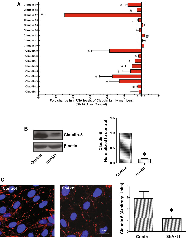
Fig. 5.
Long-term effect of Ang-1 and VEGF on endothelial-barrier protection is reliant on Akt1-mediated TJ stabilization. a Histogram showing fold-changes in mRNA levels of the claudin-family of TJ proteins with ShRNA-mediated Akt1 knockdown in HMEC, compared to control (n = 3). b Western blot showing reduced expression of endothelial claudin-5 in ShAkt1-HMEC compared to ShControl HMEC. Right panel shows a bar graph with densitometry analysis of claudin-5 bands comparing ShControl and ShAkt1 HMEC lysates (n = 3). c Representative fluorescent images showing the expression of endothelial claudin-5 in control and ShAkt1 HMEC-barrier junctions as evidenced by fluorescence immunocytochemistry. Right panel shows bar graph comparing control and SkAkt1 HMEC monolayers for claudin-5 expression in their barrier junctions as detected by immunocytochemistry and quantified using NIH-Image J software (n = 5). *P < 0.01. (Scale bar 20 µM)

