Fig. 7.
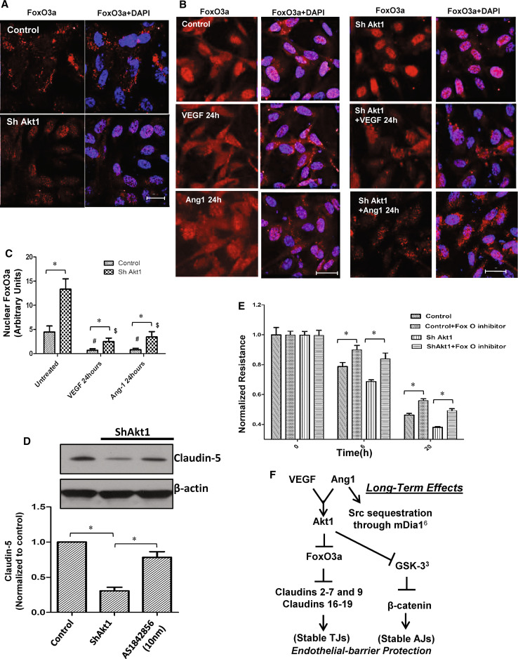
Pharmacological inhibition of FoxO transcription factors restores endothelial-barrier function in ShAkt1 HMEC. a Confocal images of ShControl and ShAkt1 HMEC stained with FoxO3a antibodies and DAPI in the presence of FBS. b Confocal images of serum starved ShControl (left panel) and ShAkt1 (right panel) HMEC, pre-treated with PBS (top), VEGF (middle) or Ang-1 (bottom) for 24 h, and stained with FoxO3a antibodies and DAPI. c Bar graph showing quantification of the nuclear FoxO3a levels from the confocal images of ShControl and ShAkt1 HMEC, pre-treated with PBS, VEGF or Ang-1 for 24 h, and stained with FoxO3a antibodies and DAPI (n = 6). d Western blot analysis of lysates from ShControl and ShAkt1 HMEC prepared after 12 h treatment with 10 nM concentration of FoxO inhibitor AS1842856 (n = 3). Bar graph showing quantification of western blot analysis of ShControl and ShAkt1 HMEC prepared after 12 h treatment with 10 nM concentration of FoxO inhibitor AS1842856 is shown below (n = 4). e Bar graph showing the effect of FoxO inhibitor AS1842856 (10 nM) on endothelial-barrier resistance in ShControl and ShAkt1 HMEC as measured from the ECIS equipment (n = 4). f Working hypothesis on the role of Akt1 on acute and chronic vascular permeability. *P < 0.01 (scale bars 20 µM)

