
Figure 1.
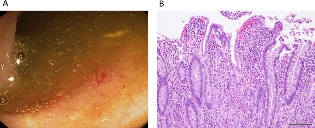
Endoscopic (A) and histologic (B) appearance of the ileum in a patient with mild UC and active ileitis. Endoscopy of the ileum (A) showed patchy erythema. Histology of the ileum (B) showed mild villous blunting, surface degenerative changes, crypt regeneration, and a mild neutrophilic infiltrate in the lamina propria and, focally, in the epithelium.