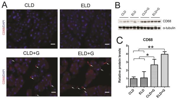
Figure 6. Ethanol-induced macrophage infiltration in the pancreas.
A: The infiltration of macrophages in the pancreas was determine by CD68 immunofluorescent staining (Red). Nuclei were labeled with DAPI (blue). Bar = 50 µm. B: The expression of CD68 in the pancreas was determined by immunoblotting. C: The relative expression of CD68 was quantified and normalized to α-tubulin. Each data point was the mean ± SEM of three animals. * denotes statistical difference (p<0.05) and ** denotes significant difference (p<0.01).

