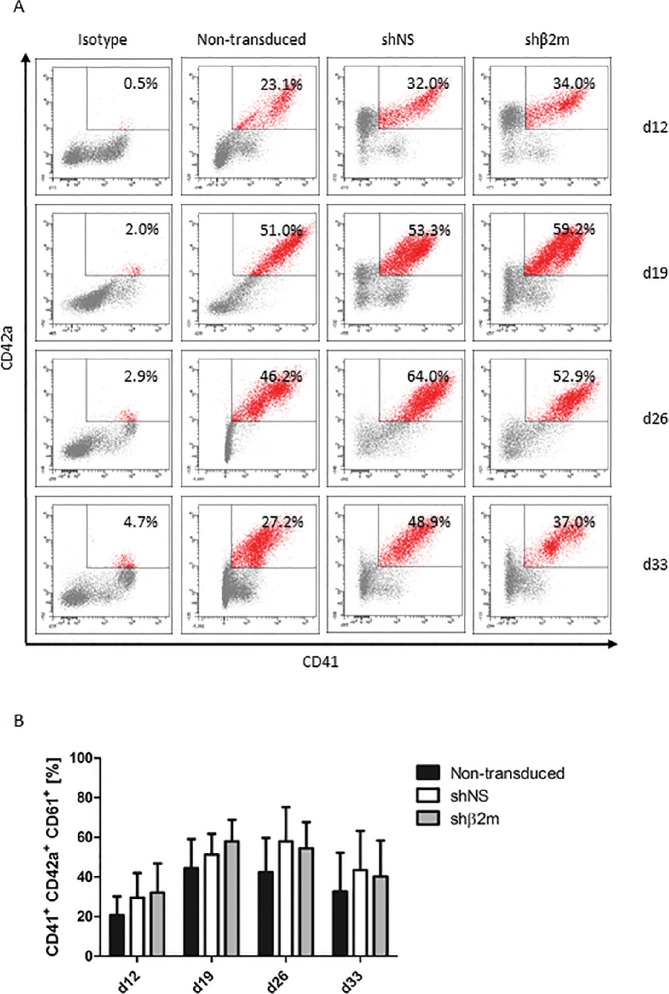
Figure 3.
Characterization of iPSC-derived HLA class I-silenced megakaryocytes (MKs). Nontransduced iPSCs and iPSCs expressing either a control nonspecific shNS or a shRNA targeting β2m transcripts (shβ2m) were cultured for 33 d and analyzed weekly from d 12 for MK differentiation (four time points). iPSCs were transduced with shRNA encoded in a pLVTHm backbone. (A) MKs were identified based on the expression of CD41 (GPIIb) and CD42a (GPIX). Representative flow cytometry dot plots are shown for each condition and time point. (B) Mean and SD of CD41+CD42a+CD61+ cell frequencies were detected by at least four independent experiments. (C) Polyploidy analysis of shNS or shβ2m-expressing CD41+ cells on d 26 and d 33. Representative flow cytometry histograms are shown. (D) Representative fluorescence microscopy images of MKs exhibiting polyploid nuclei (lower row) and morphological analyses of proPLT-forming MKs (upper row). Representative pictures of nontransduced, shNS or shβ2m-expressing MKs on d 20 of differentiation are shown (bright field).

