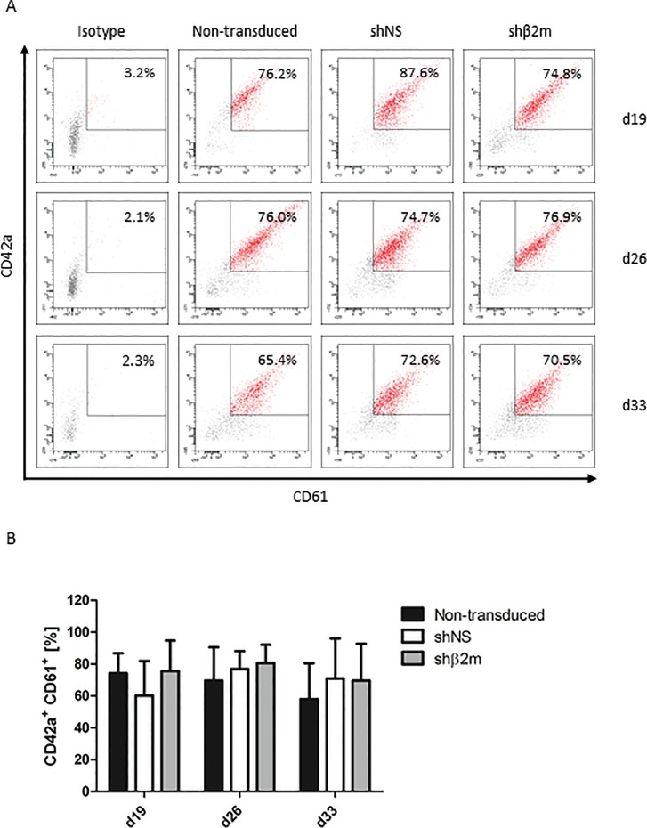
Figure 5.
Characterization of HLA class I-silenced iPSC-derived platelets (PLTs). Nontransduced iPSCs and iPSCs expressing either a control nonspecific short-hairpin RNA (shNS) or a shRNA targeting β2-microglobulin transcripts (shβ2m) were cultured for 33 d and analyzed weekly from d 12 for PLT differentiation (four time points). (A) PLTs were identified by selection of a CD41+ (GPIIb) population. Shown is the percentage of CD42a+ (GPIX) CD61+ (GPIIIa) co-expression of CD41+ cells. Representative flow cytometry dot plots are shown for each condition and time point. (B) Mean and SD of CD42a+CD61+ cells of four independent experiments.

