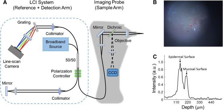
FIG. 1.
A Schematic of the combined LCI-otoscope imaging system used in the study. B TM surface image, similar to a video-otoscope image, acquired by the CCD camera. C Depth-resolved LCI data acquired at the imaging site marked by the red point in (B). The two prominent peaks correspond to the epidermal and the mucosal surfaces of the TM. TM thickness is estimated as the distance between the two peaks.

