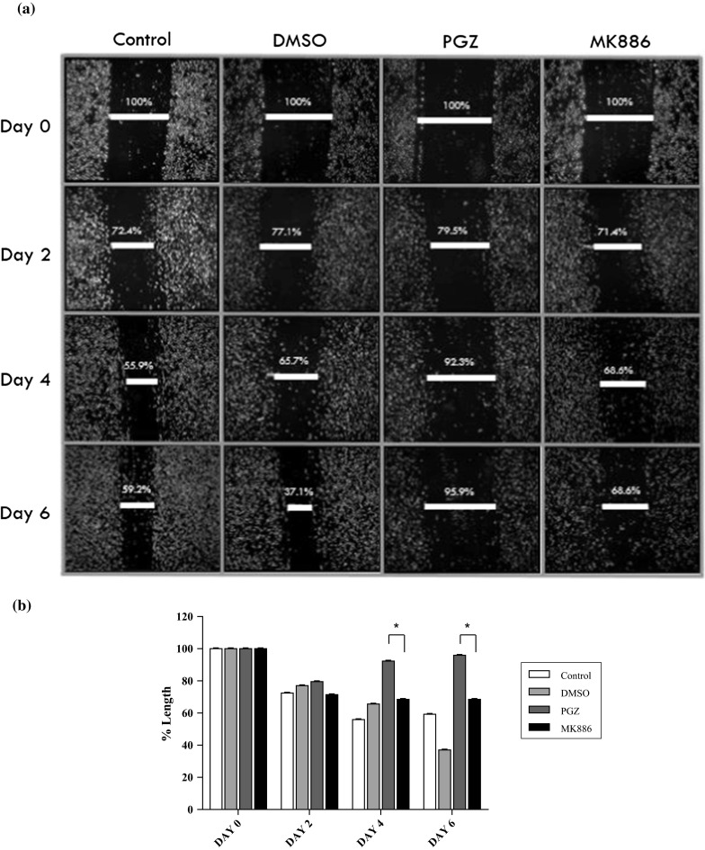
Fig. 5.

a The invasion of PGZ-treated and MK886-treated MDA-MB-231 cells as verified by the in vitro wound healing migration assay. Cell migration was estimated by measuring the gap within the wounded region. b Statistical analysis of the PGZ-treated and MK886-treated MDA-MB-231 cells for 2, 4 and 6 days. Data are shown as the mean ± SD of triplicate cultures, *p < 0.05
