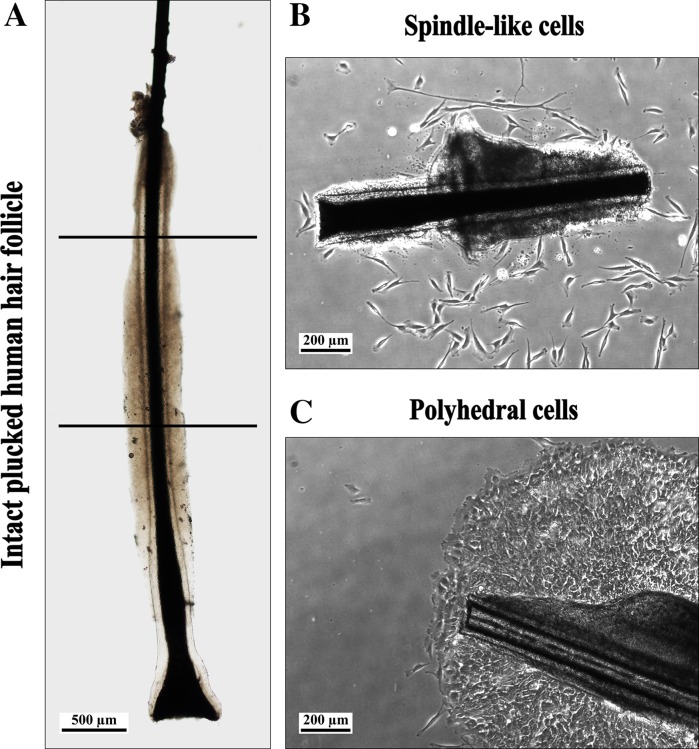
Fig. 1.
a Hair follicle with an intact inner and outer root sheath. Only the upper half of the follicle was used (between lines; scale bar 500 µm). b HF and cells with spindle-like morphology, at day 2 of outgrowth. The outer root sheath is curled (scale bar 200 µm). c HF and tightly clustered cells with an epithelial appearance (sheets of flattened polyhedral cells; scale bar 200 µm)

