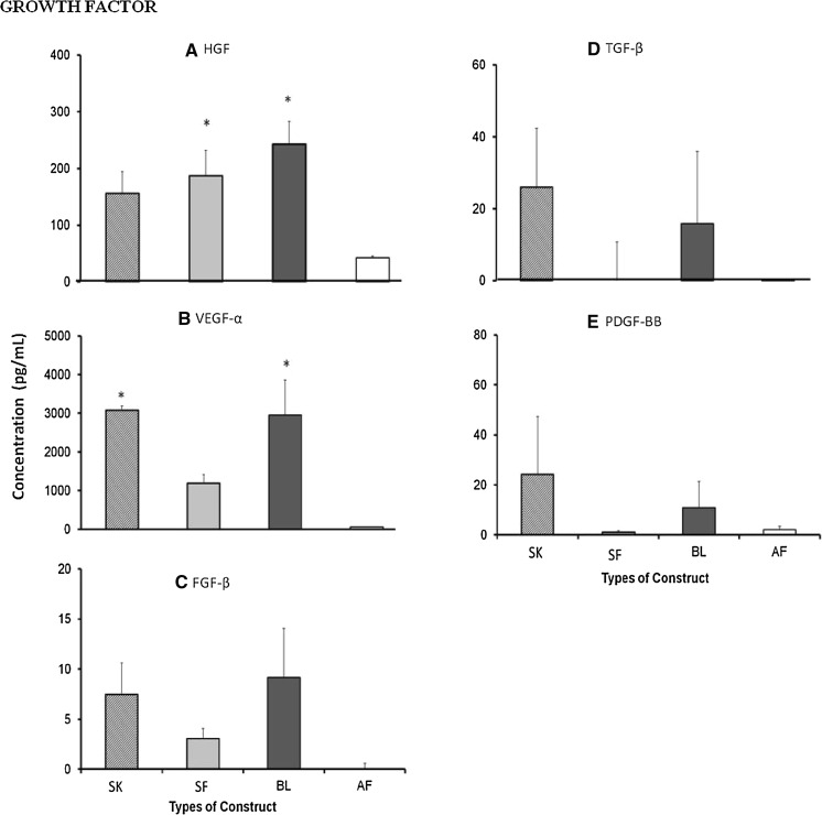
Fig. 4.
Wound-healing growth factors (a HGF, b VEGF-α, c FGF-β, d TGF-β, e PDGF-BB) secreted by single layer keratinocytes (SK), single layer fibroblasts (SF), bilayer (BL) and acellular fibrin (AF) substitutes. No significant difference was observed for secretion of growth factor HGF, VEGF, FGF-β, TGF-β and PDGF-BB for all three substitutes SK, SF and BL. Asterisk represents significantly higher compared to AF substitute (p < 0.05)

