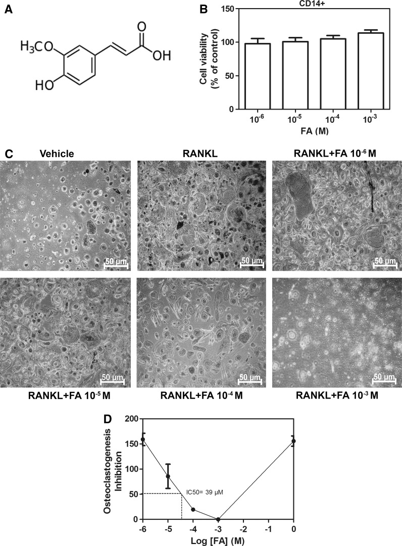
Fig. 1.
Effects of FA on cell viability and osteoclast differentiation. a Molecular structure of FA. b Cell viability of FA-treated human CD14+ monocytes. Cells were treated with indicated concentrations of FA for 48 h and cell viability was measured by alamar blue assay. c CD14+ monocytes were treated with RANKL in the presence or absence of FA and differentiated into osteoclasts as mentioned in methods and TRAP staining for osteoclasts was performed. d Graph representing inhibition of osteoclast formation by FA. Relative IC50 was calculated by nonlinear regression log (FA concentration) versus response–variable slope. Data are expressed as mean ± SD and are representative of three independent experiments

