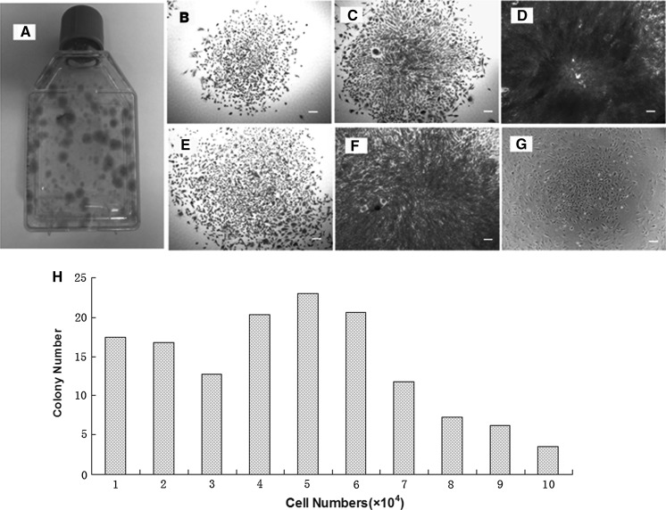
Fig. 1.
The colony formation of meniscus-derived stem cells (MMSCs) and quantitative analysis of colonies. a Colonies of MMSCs were detected in one of 5 T25 flasks by staining with Methyl violet at 15 days. b–f Five distinct colony types were visualized morphologically in rabbit meniscus-derived stem cells cultured either in T25 flasks (b, c, e) or 96-well plates (d, f). g Lots of colonies were still observed when cultured at passage 5 despite their decreasing numbers compared to passage 1. h Cell numbers were counted after trypsinized from each colony respectively (magnification of microscopy: ×4) (Bar 50 μm)

