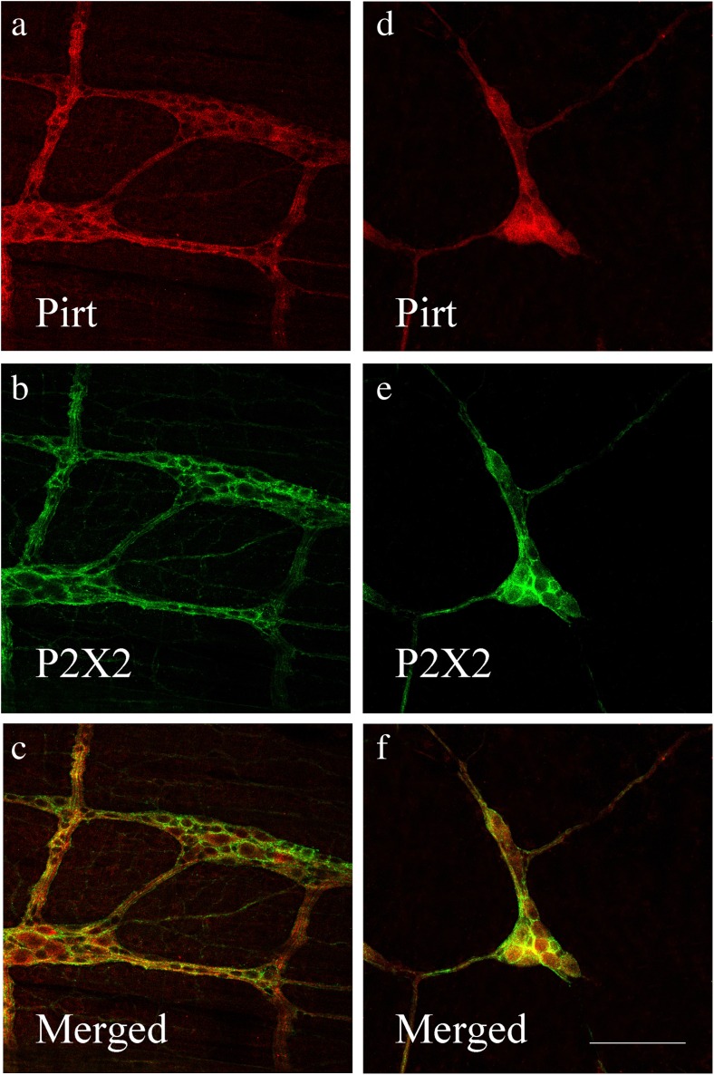
Fig. 8.
Co-localization of Pirt-ir and P2X2 receptor-ir cells in the myenteric plexus (a–c) and submucous plexus (d–f) of the mouse ileum (by using confocal microscopy). a Pirt-ir cells (red) in the myenteric plexus. b P2X2-ir cells (green) in the myenteric plexus from the same section as a. c Merged image from a, b. Double-labeled cells are yellow in color. d Pirt-ir cells (red) in the submucous plexus. e P2X2-ir cells (green) in the submucous plexus from the same section as d. f Merged image from d, e. Double-labeled cells are yellow in color. Scale bars, a–f 250 μm

