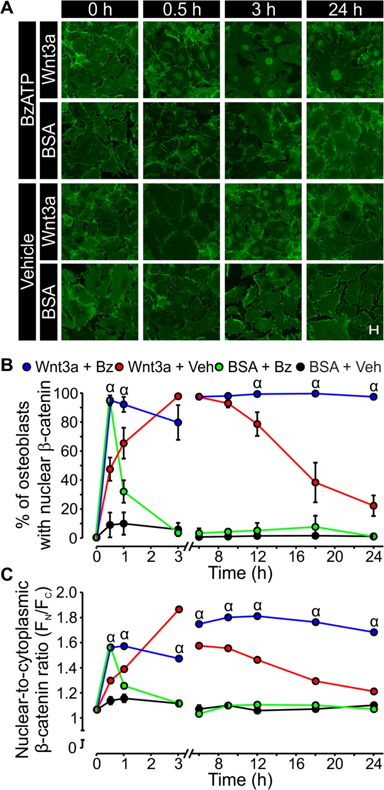
Fig. 1.
The P2X7 agonist BzATP potentiates β-catenin nuclear localization elicited by Wnt3a. a MC3T3-E1 osteoblast-like cells were seeded at a density of 1.5 × 10⁴ cells/cm2 on glass coverslips and cultured for 2 days. Cells were then placed in serum-free media and incubated overnight. The next day, cells were treated under serum-free conditions with recombinant mouse Wnt3a (20 ng/ml) or its vehicle (0.2 % BSA; BSA) in the presence of either BzATP (300 μM) or its vehicle (Vehicle or Veh) and fixed at the indicated times. To observe changes in subcellular localization of β-catenin, immunofluorescence was performed using a β-catenin monoclonal antibody (green) and visualized by confocal microscopy. Data are representative images from four independent preparations each performed in duplicate. Scale bar is 20 μm. b β-catenin nuclear localization was assessed by measuring the average pixel intensity of an area in the nucleus (F N) and an area of equal size in the cytosol (F C). The proportion of all cells within 24 fields (12 per coverslip) exhibiting β-catenin nuclear localization was analyzed for each condition. Values of the ratio F N/F C greater than or equal to 1.25 were taken as indicating nuclear localization. The number of cells exhibiting nuclear localization was expressed as a percentage of the total number of cells for each treatment group. c The ratio of F N/F C (an indication of the intensity of β-catenin nuclear localization) for all cells analyzed was plotted for each treatment at times indicated. For both b and c, α indicates a significant effect of BzATP on Wnt3a-induced β-catenin nuclear localization compared to Wnt3a alone at the same time point (p < 0.05). Data are means ± SEM (n = 4 independent preparations each performed in duplicate)

