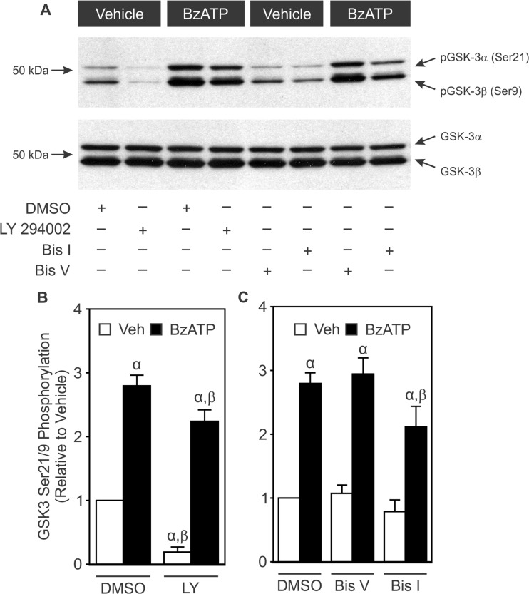
Fig. 8.
Effects of PI3K and PKC inhibitors on BzATP-induced inhibitory phosphorylation of GSK3α/β. Changes in inhibitory phosphorylation of GSK3α/β were monitored in MC3T3-E1 cells as described in the legend to Figure 6. Cells were incubated for 20 min in the presence or absence of the PI3K inhibitor LY 294002 (30 μM; LY), its vehicle (DMSO), the PKC inhibitor bisindolylmaleimide I (0.5 μM; Bis I), its inactive analogue bisindolylmaleimide V (0.5 μM; Bis V), or their vehicle (DMSO). Next, cells were treated with vehicle (Veh) or BzATP (300 μM; BzATP) for 5 min, and total protein was subsequently harvested. Samples were subjected to Western blot analyses using a specific pSer21/9 GSK3α/β antibody. As a loading control, blots were also probed for total GSK3α/β. Bands were visualized by the ECL method. a Immunoblot representative of three independent preparations. b, c pSer21/9 and total GSK3α/β bands were evaluated by densitometry. The ratio of pGSK3α/β to total GSK3α/β was determined, and results were normalized to Veh + DMSO. α indicates a significant difference from Veh + DMSO (p < 0.05). β indicates a significant effect of the inhibitor (p < 0.05). Data are means ± SEM (n = 3 independent preparations)

