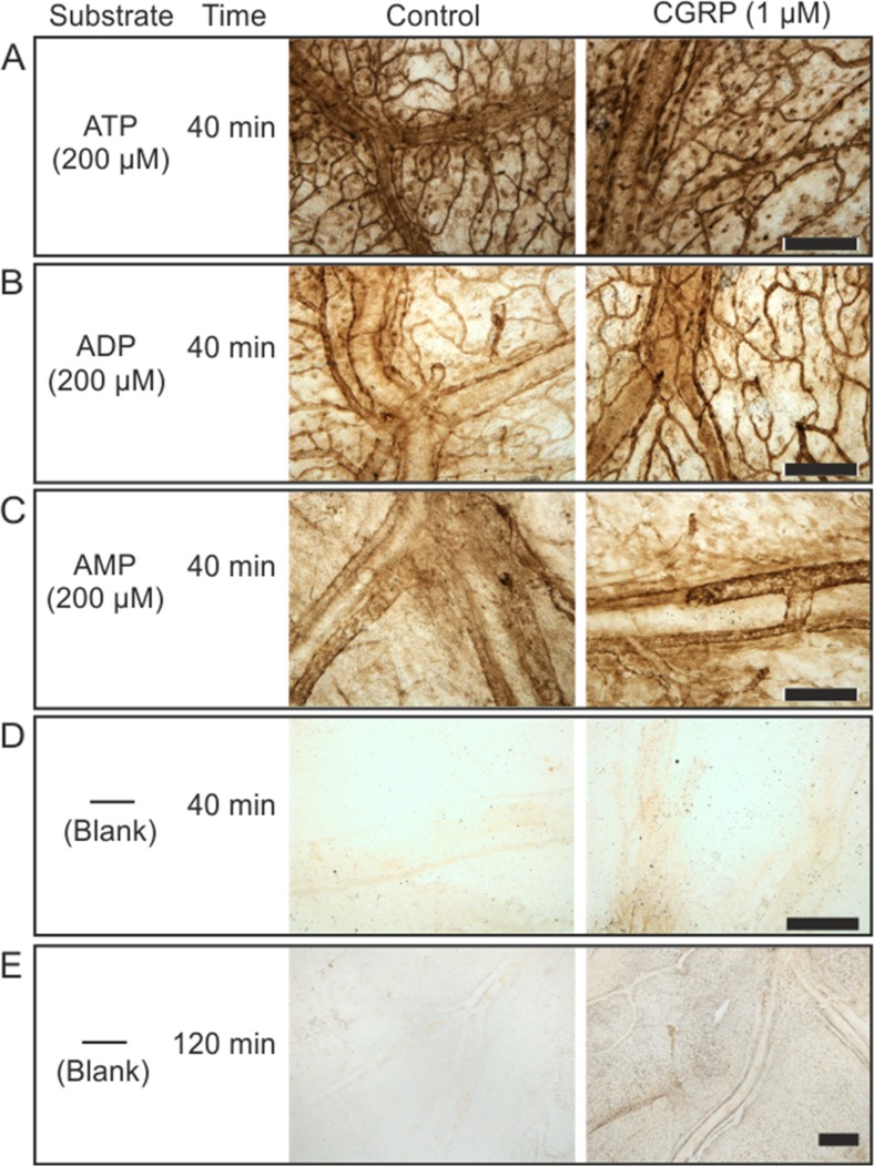
Fig. 3.
Histochemical analysis of distribution of nucleotidase activities in whole mount rat meninges. Rat hemiskulls were pre-treated for 2 h without and with 1 μM CGRP and subsequently subjected to enzyme histochemical staining by incubating the meninges for 40 min with 2 mM Pb(NO3)2 and 200 μM of one of the following nucleotides: ATP (a), ADP (b), and AMP (c). Representative staining of control and CGRP-treated meninges are also shown after their incubation for 40 min (d) and 2 h (e) with medium containing only lead nitrate, without any exogenously applied nucleotide substrate. Strikingly, increasing the incubation time revealed clear-cut increases in blank meningeal staining after tissue exposure to CGRP. Scale bars, 300 μm

